


Progress in Nanotechnology and Development of a New Medical Imaging System
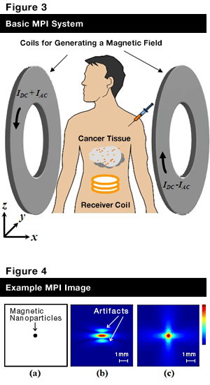
There has been a great deal of research on magnetic nanoparticles*1 recently, serving to create innovative materials and making a significant contribution to the development of new technologies and systems in various fields. In the medical field, in particular, a wide variety of applications are envisioned for magnetic nanoparticles, and they are already used as a contrast agent in magnetic resonance imaging (MRI) to construct a clear image of the lesion location. In 2005, a magnetic particle imaging (MPI) technique was proposed for early detection of disease, including cancer, which leverages the unique properties of magnetic nanoparticles.Principle – How MPI Works
Nanoparticles, having the diameter of several tens of nanometers, accumulate in cancerous tissues. This is attributable to abnormal angiogenesis*2 induced by rapid proliferation of cancer cells. Abnormal angiogenesis creates a vascular structure with gaps one-digit wider than in normal blood vessels, usually in several hundreds of nanometers, through which nanoparticles seep into cancerous tissue and stay there for a specific period of time. Identifying the location of these magnetic nanoparticles seeped into cancerous tissue enables construction of an image and early detection of cancer.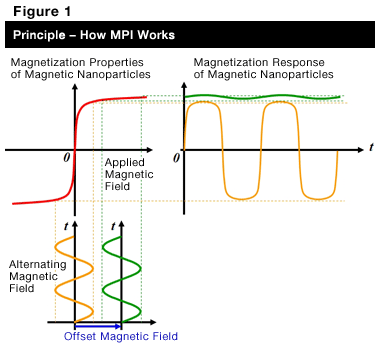
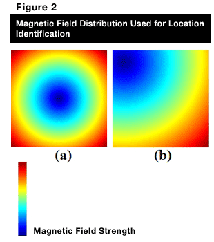
Magnetic nanoparticles show magnetic behavior (magnetization) (red line in Figure 1) when exposed to a magnetic field. Applying an alternating magnetic field also makes the magnetization intensity of nanoparticles to vary over time (orange line in Figure 1). Application of another strong magnetic field (an offset magnetic field) to these nanoparticles makes the intensity of magnetization remain constant (saturation of magnetization) with no additional changes in intensity observed over time (green line in Figure 1). Figure 2 (a) shows a magnetic field distribution of varying strengths (weak in one area and stronger in the surrounding areas). By applying an alternating magnetic field to this magnetic field distribution while scanning its spatial data, as depicted in Figure 2 (b), and by detecting variations in magnetization of nanoparticles, we can determine whether these nanoparticles exist in the area with low field strength. This, in turn, allows construction of an image of the area in which magnetic nanoparticles accumulate, i.e., the location of cancer.
Current Status of MPI
Future of MPI Research on MPI has been conducted by research groups in Germany (University of Lübeck、Royal Philips Electronics, Bruker Corp. and others), University of California, Berkeley in the United States and Meiji University in Japan. Studies on image construction methods and system building have just begun, and much remains to be solved. Some have noted, however, that the current situation is very similar to what had happened in the early stage of MRI development, and there is a possibility that development of MPI may proceed at a considerable pace in the future. Moreover, generating heat in magnetic nanoparticles accumulated in cancerous tissues could heat up and kill these tissues, and we are simultaneously accelerating research on a system for magnetic hyperthermia. We hope to create a system that fuses diagnostic and treatment functions for detection and treatment of cancer in an extremely early stage, causing less burden on patients.
*1) Magnetic nanoparticles: Particles at the nanoscale, which show magnetic behavior when exposed to an external magnetic field. (One nanometer is equal to one billionth of a meter.)
*2) Angiogenesis: A phenomenon encouraging growth of new blood vessels in cancer cells to take in oxygen and nourishment. Growth of new blood vessels is controlled in normal cells.
Profile
Associate Professor of Department of Mechanical Engineering Informatics, School of Science and Technology. Ph.D. (Engineering)Born in Aichi Prefecture in 1964. Graduated from Department of Electrical and Electronic Systems Engineering, School of Engineering, Nagaoka University of Technology in 1987. Completed master’s course in Department of Electrical and Electronic Systems Engineering, Graduate School of Engineering, Nagaoka University of Technology in 1989. Worked for the Research and Development Center (renamed Corporate Research and Development Center) of Toshiba Corporation from 1989 to 1999, the Medical Systems Company of Toshiba Corporation from 1999 to 2003 and Toshiba Medical Systems Corporation from 2003 to 2004. Worked as Associate Professor from 2004 to 2010 in Department of Electrical Engineering, Nagaoka University of Technology. Assumed current position in 2010.
Main Publications (Co-Authoring)
Non-invasive Localized Heating and Temperature Monitoring Based on a Cavity Applicator for Hyperthermia, New Developments in Biomedical Engineering, Domenico Campolo (Ed.), pp. 569 - 590,ISBN: 978-953-7619-57-2, InTech, 2010Image Resolution and Sensitivity Improvements of a Molecular Imaging Technique Based on Magnetic Nanoparticles, Electromagnetic Waves, Vitaliy Zhurbenko (Ed.), pp. 493 - 510,ISBN: 978-953-307-304-0, InTech, 2011
Academic Societies
Japanese Society for Medical and Biological EngineeringJapanese Society for Magnetic Resonance in Medicine
The Institute of Electronics, Information and Communication Engineers
Japanese Society for Thermal Medicine
The Society of Instrument and Control Engineers


